Application step by step
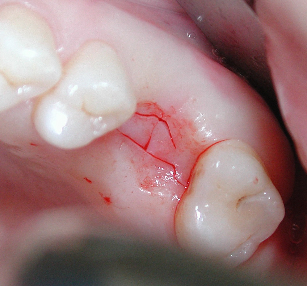
1. Crestal longitudinal incision and mucoperiostal flap opening

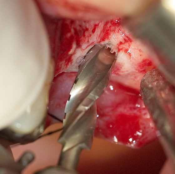
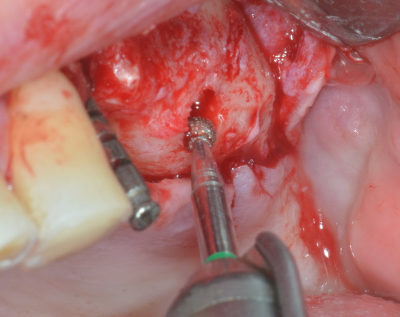
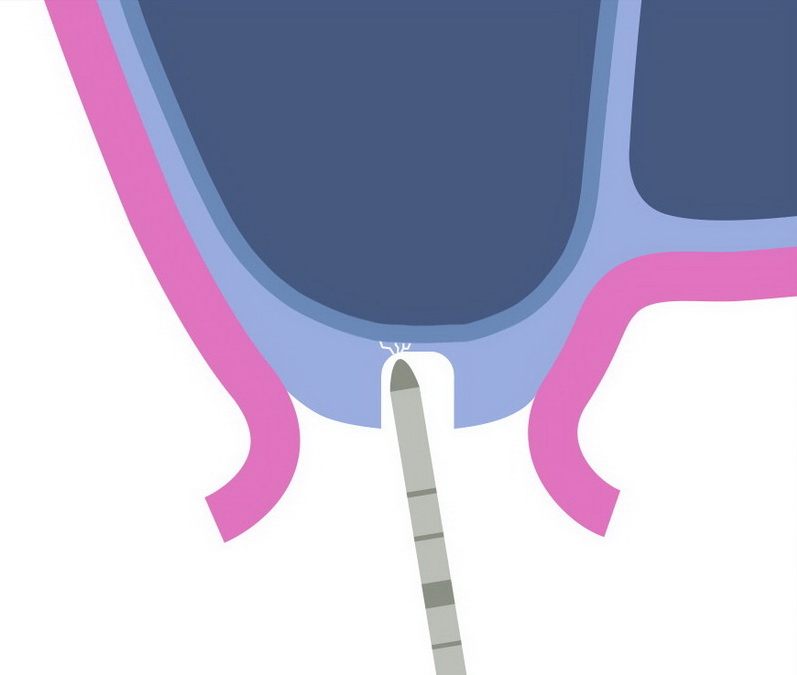
2. Implant drilling down to 1 - 2 mm above the sinus floor
Starting with an used pilot bur with poor grinding capacity in order to feel better the sinus corticalis

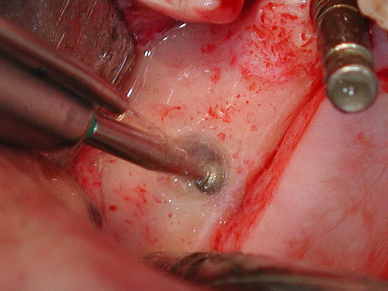
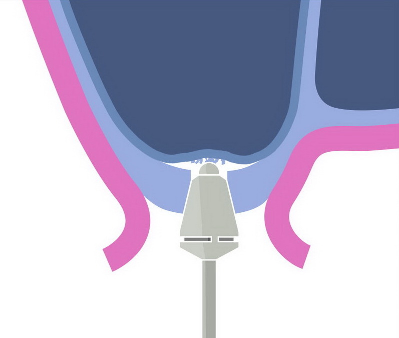
3. Thinning of the sinus corticalis with a round diamond bur

At a residual bone height of 3 mm or less the diamond drill makes an opening without any prior implant drilling
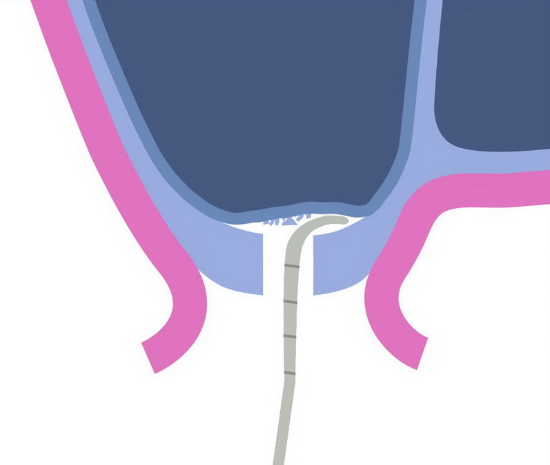
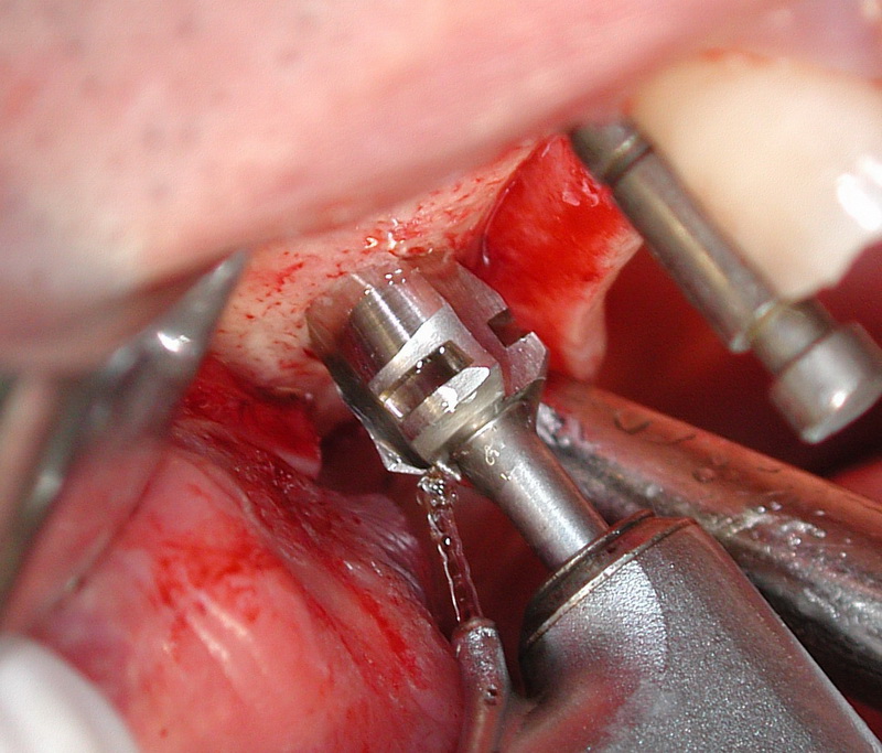
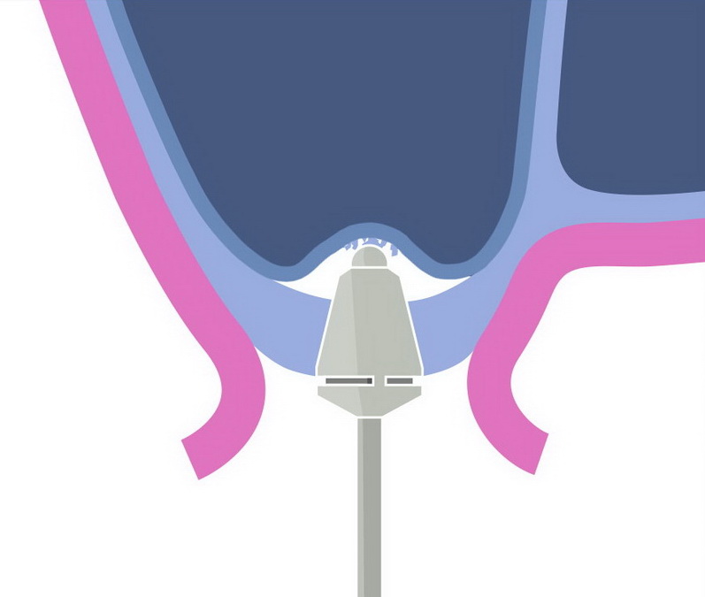
4. Three to four marginal predetermined breaking points are made with a fine, convex osteotome.
5. Breaking through the sinus floor, penetrating at most 1 - 2 mm into the sinus, first with the convex osteotome…

… then with the concave osteotome.
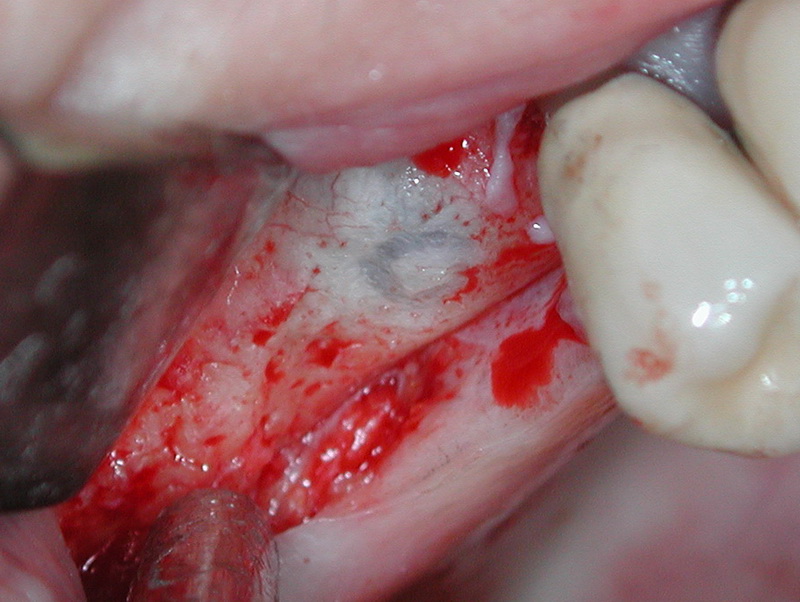
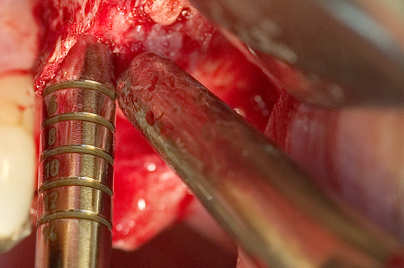
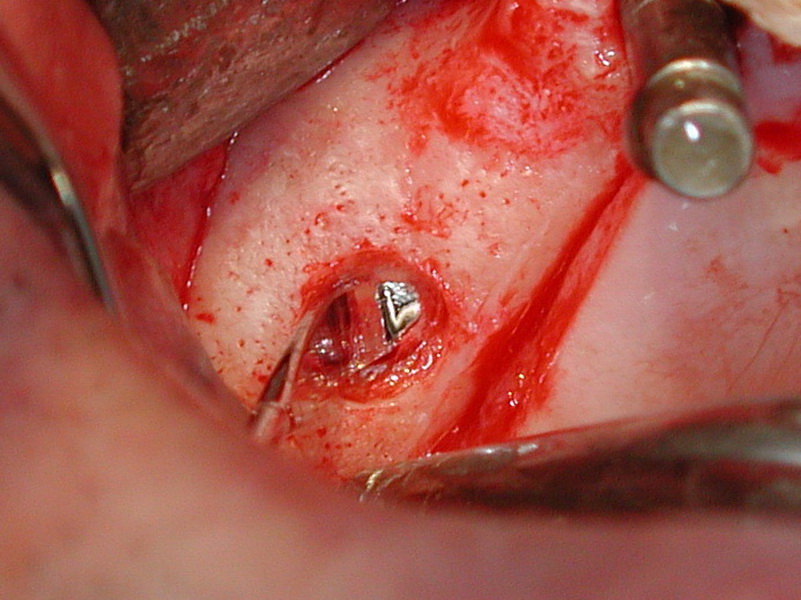
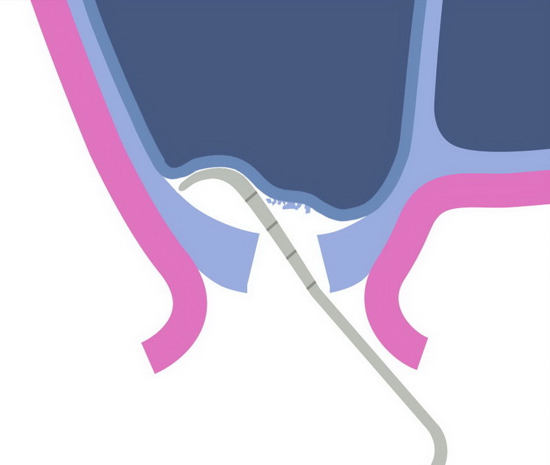
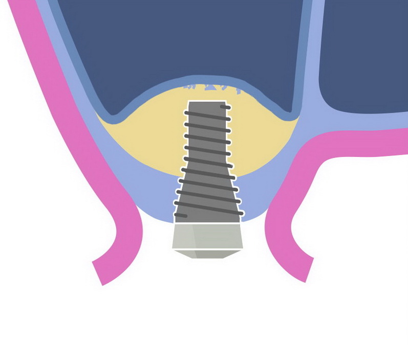
6. Circular detachment and lifting of the sinus mucosa with the Benex sinus elevators to about five millimeters
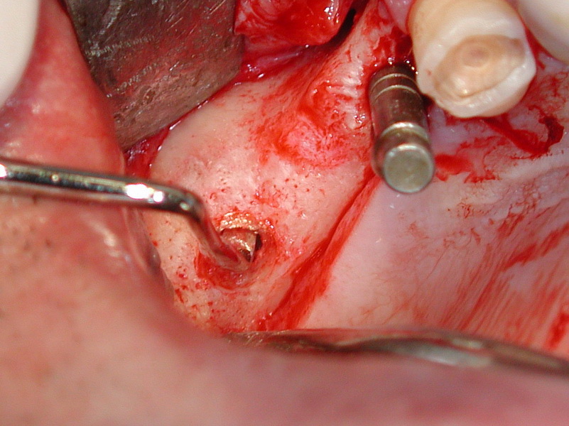
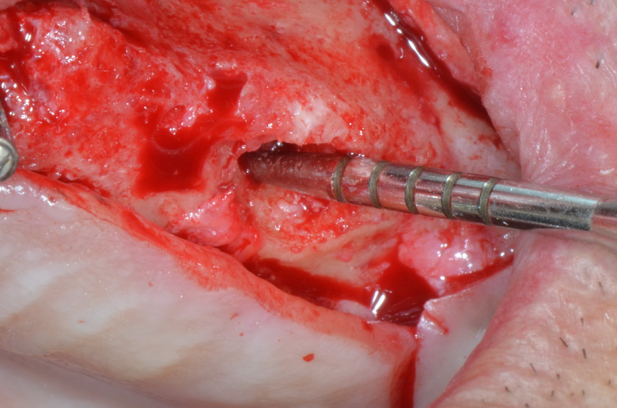
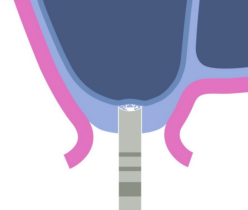
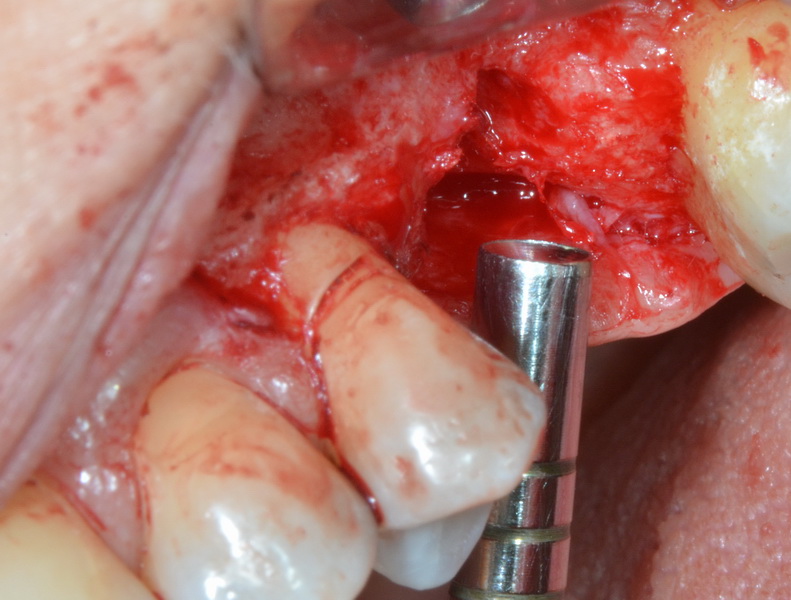
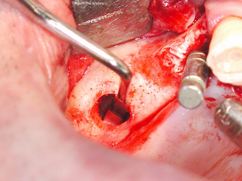
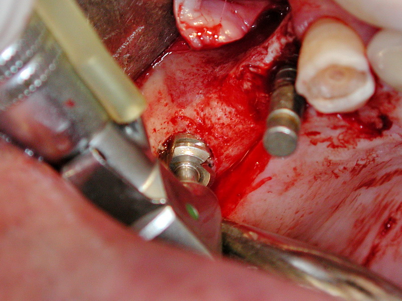
7. Widening of the implant bed with profile drill.
The business end of the drill must be rounded.
8. Further elevation of the sinus mucosa with the Benex elevators up to the desired implant length
9. Profile drilling for the implant's end position
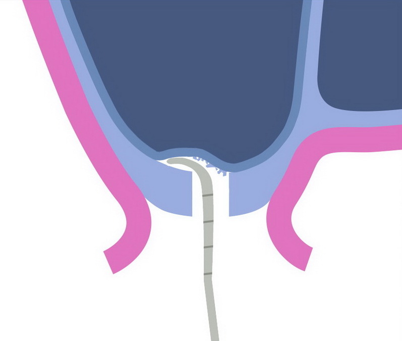
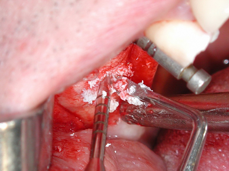
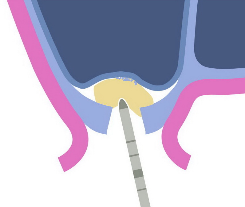
10. Vibrating, the bone substitute is introduced under the sinus mucosa with the finest, convex osteotome.
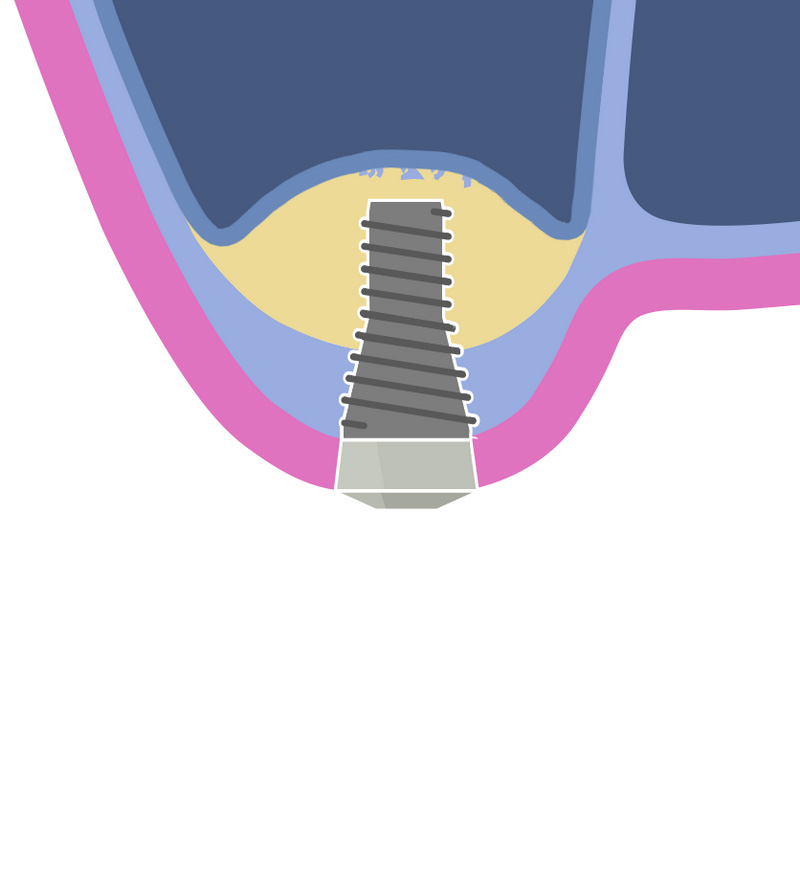
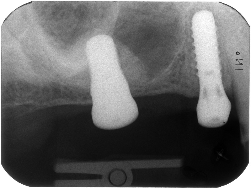
Insertion of the coronal-tapered implant (X-ray postop)
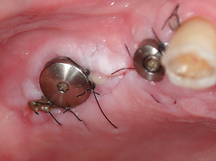
Wound closure (situ 10 days postop)